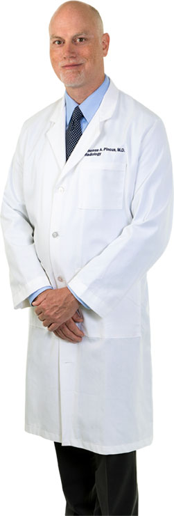
Peninsula Radiological Associates; Medical Director Radiology, Riverside Regional Medical Center
 When Dr. Thomas Pincus was a medical student two decades ago, many of the patients he treats today would have had no chance of survival – or at least not without devastating health complications.
With dramatic advances in imaging technologies, rapid stroke interventions and radiation therapies, Dr. Pincus has seen his subspecialties of Neuroimaging and head and neck imaging evolve rapidly. He also has helped guide the growth of Riverside Regional’s neurosciences program into the Comprehensive Stroke Center it is today.
Yet to Dr. Pincus, the most rewarding part of being a physician is helping put his patients at ease on a daily basis, guiding them through common-yet-frightening procedures such as biopsies, drainages, barium swallows and MRIs.
“All physicians are busy, but the bottom line is we’re always treating a human being,” he says. “I like taking the extra time to explain what we plan to do together, step by step. My goal is to support them through a difficult time with as little discomfort and anxiety as possible, and with a high degree of safety.”
While always keeping the big picture in mind, Dr. Pincus also encourages his patients to take each phase of treatment as it comes: “Otherwise, it’s too easy for them to get overwhelmed and discouraged.”
Not surprisingly, Dr. Pincus has been named a Hampton Roads “Top Doc” by Coastal Virginia Magazine five times and is a popular lecturer for medical residents. He also is a respected leader at his practice as well as at Riverside, where he directs weekly neuroscience case conferences and has served on the hospital’s stroke, MRI safety and surgical quality and performance committees.
A Hampton native, Dr. Pincus grew up as the third youngest of 12 siblings. By the time he was a student at Hampton High School, he had decided to follow an older brother, an ENT physician, into medicine.
After majoring in biology at the University of Dallas, Dr. Pincus earned a medical degree from University of Texas Health Science Center at Houston. He completed a general radiology residency and a neuroradiology fellowship at University of Texas Medical Branch at Galveston.
Dr. Pincus has worked with Peninsula Radiological Associates since 2002. Board certified by the American Board of Radiology, he holds medical licenses in Virginia, North Carolina and Texas and enjoys collaborating with his partners and a variety of specialists in the region.
“Radiology allows for interaction with many fields of medicine, and I’ve always found the anatomy and function of the brain and spine especially interesting,” he says. “The ability to use MRI and CT to clearly see the type of pathology and plan treatments is very powerful.”
CT angiography, for example, is a critical part of diagnosing and treating ischemic or hemorrhagic stroke. Scans taken within 15 minutes of a patient’s arrival at Riverside’s emergency room now can clearly image blood vessels from the top of the heart into the brain, including the carotid and vertebral arteries, in isolation from other structures.
That ability to precisely visualize blockages, blood clots and arterial damage, as well as unexpected findings such as tumors or lesions, has led to much quicker decisions on effective interventions.
“We are seeing very good outcomes – limited damage or complete resolution – for what easily could have been a devastating or fatal stroke,” Dr. Pincus notes.
Many brain cancer patients, meanwhile, qualify for gamma knife therapy, a minimally-invasive procedure to pinpoint and often destroy metastatic lesions without remissions. That can eliminate repeated doses of whole brain radiation, a threat to mental capacity over time.
Dr. Pincus’ job is to perform and interpret MRI scans before neurosurgeons and radiation therapists take over. “By quantifying and identifying the exact locations of cancerous lesions to target, we can save patients we couldn’t save in the past,” he states. Emerging chemotherapy methods that deliver drugs directly to tumors will rely on such detailed imaging, too.
The future looks promising for even more types of patients. MR magnets, for instance, can produce images of spinal tracts, or the pathways for sensory or motor nerve impulses within the spinal cord’s white matter.
“We now can determine where nerve tracts are disrupted, or if a tumor is very nearby,” Dr. Pincus says. “That gives neurosurgeons an amazing capacity to remove tumors without disturbing nerve tracts, or for alternatives such as radiosurgery to prevent sensory or motor/muscular damage.”
In head and neck imaging, modalities such as PET/CT, CT perfusion/angiography and Diffusion MRI aid in cancer diagnosis and staging, along with rapid evaluation of infections and traumatic injuries to the head and neck. In nuclear medicine, improvements in radiotracers used in PET scans may be helpful in identifying patients with Alzheimer’s and Parkinson’s diseases sooner.
For cancer patients, minimally-invasive ablation of tumors with heat, electrical energy or extreme cold is providing an alternative for people who don’t qualify for surgery. Again, imaging tests are critical to properly positioning a needle probe into a tumor.
Outside the office, Dr. Pincus has been married for 13 years to wife Stephanie and has five children, ages 2 through 11. His hobbies include surfing, fishing and playing tennis.
Dr. Pincus remains committed to developing local neuroimaging capabilities to give his patients the best chance of a positive outcome. All the while, he’ll be an advocate by their side.
“The less people understand about what’s happening to them, the more scared they will be,” he says. “They’re not used to being in a hospital, and they deserve to have my time and encouragement. They always deserve to know what to expect.”

