
Comprehensive Neurosciences:
exceptional neurovascular and neurosurgical care for all of Hampton Roads
Unexplained weakness. Pain. Tremor. Numbness. Blurry vision. Headache. These can be among the most bothersome symptoms a patient can experience. They’re also among the symptoms that patients frequently ignore, believing they’ll resolve on their own. Indeed, they often do.
But these symptoms can also be indicators of dangerous neurological conditions, which, if left untreated, can have devastating and even lethal consequences. When such symptoms linger, the patient should be immediately referred to a neurological specialist for a thorough evaluation and diagnosis – and given the opportunity to consider appropriate conservative treatment.
When conservative treatment fails or is inappropriate, patients and referring physicians throughout Hampton Roads need look no further than Riverside Comprehensive Neurosciences for exceptional care by world-class physicians, who have mastered the exacting and delicate technology available to treat neurological illness and injury.
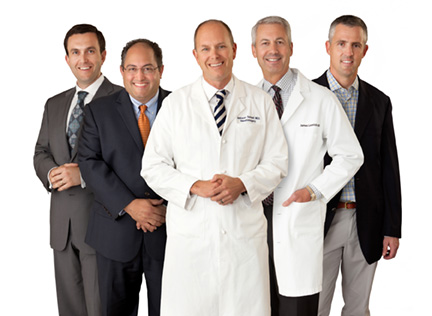
Riverside Comprehensive Neurosciences has expanded exponentially, boasting the newest, state-of-the-art technology for treating neurological illness and injury. Riverside Comprehensive Neurosciences offers patients and their referring physicians the combined expertise of a team of fellowship-trained neurosurgeons, interventional neuroradiologists, neurologists, medical physicists, radiation oncologists and other highly skilled professionals. Each individual patient receives the full benefit of their collective body of knowledge, training and experience.
Unprecedented precision when micro-measurements count.
From the simplest to the most complex case, Riverside Comprehensive Neurosciences offers treatment modalities and care equivalent to the largest medical centers in the country. The use of a bi-plane digital x-ray in treating aneurysm cases is one example.
Statistics show that as many as 3 to 5 percent of people have one or more of these small, slow growing ‘bulges’ that form at weak spots in the wall of a blood vessel in the brain. Because in their earliest stages, aneurysms are asymptomatic, often the first sign of trouble is when they rupture. Between 30 and 50 percent of patients with a ruptured aneurysm die, or are left with significant disability; but when patients receive appropriate care in a timely fashion, they can survive and even thrive.
Timely treatment used to mean a craniotomy – a difficult surgery that involves removing the skull, retracting the brain and clipping the aneurysm.
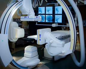
Bi-plane digital x-ray.
As explained by neuroradiologist Frank Sanderson, MD, the procedure is far less invasive; it’s safer and much easier on the patient. Utilizing the bi-plane digital x-ray for guidance, Riverside surgeons insert a flexible catheter into the femoral artery, and thread it up through the neck into the brain. The bi-plane utilizes two mounted, rotating cameras – one on each side of the patient, taking simultaneous pictures that are fed into a computer, which takes simultaneous two-dimensional images and creates a high-resolution 3-D reconstruction of the aneurysm. The surgeons can manipulate the image to give them an absolute, perfect projection of the aneurysm. They can then insert a smaller catheter into the aneurysm through which progressively smaller platinum coils can be introduced until the aneurysm is tightly packed, thus depriving the aneurysm of its blood supply. The patient, now headache and symptom free, goes home the next day.
The extraordinary high-resolution visualization of the brain’s vascular network made possible by the imaging technology is assisting surgeons in managing stroke cases as well. With a fellowship-trained stroke neurologist, Wolfgang Leesch, MD, a neuroradiologist and a neurosurgeon on hand, stroke victims get the benefit of three different perspectives. The images revealed on the computer screen help surgeons guide catheters directly to the clot with pinpoint accuracy, allowing its removal from the brain. The formerly ominous empty space on x-ray, once blocked by the clot, is filled with vasculature as blood flows freely again.
It’s important to note, however, Dr. Leesch emphasizes, that results always depend on how much time passes between the blockage and the time it’s opened. Many more lives could be saved if patients could learn to act quickly.
A true Center of Excellence.
In 2006, understanding the potential of the stereotactic surgery being done by pioneering practitioners at the University of Virginia, Riverside Comprehensive Neurosciences partnered with UVA Health System to bring the technology to the Virginia Peninsula. James Lesnick, MD was instrumental in putting this partnership together. Today, he serves as co-medical director with Dr. Jason Sheehan from UVA.
In 2012, Chesapeake Regional Medical Center joined the alliance, and the Chesapeake Regional, Riverside & University of Virginia Radiosurgery Center was created.
Stereotactic radiosurgery – the gold standard for brain tumors and abnormalities.
The concept of stereotactic radiosurgery, introduced in 1951 by Dr. Lars Leksell, has an impressive track record in treating brain tumors and other abnormalities. Dr. Leksell understood that the nervous system doesn’t tolerate radiation as well as other parts of the body; thus his discovery has led to methods of limiting the toxicity of radiation to normal healthy tissue, while maximizing the amount of radiation that can be given to malignant or neoplastic tissue.
No other non-invasive treatment method in the field has greater clinical acceptance anywhere in the world. The Radiosurgery Center employs two modalities that deliver stereotactic radiosurgery, both of which focus very high beams of radiation on a small part of the body. Dr. William H. McAllister, IV, a neurosurgeon, explains the difference between the two modalities.
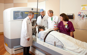
Gamma Knife.
The Gamma Knife is used for patients with brain tumors as well as other conditions, such as arteriovenous malformations, trigeminal neuralgias or vestibular schwannomas (acoustic neuromas.) Often mistaken by patients as utilizing actual surgical knives making incisions, the Gamma Knife in actuality is 201 beams of radiation all coming on at the same time and meeting at one very carefully calculated point in the brain. Invented in 1967 by Dr. Leksell with Drs. Ladislau Steiner and Borje Larsson, the Gamma Knife administers high-intensity cobalt radiation therapy that concentrates the radiation over a small volume. Members of the professional staff at the Radiosurgery Center were fortunate enough to cultivate a working relationship with Dr. Steiner during his tenure as head of the Lars Leksell Gamma Knife Center at the University of Virginia, and bring to bear the benefits of that consultation for their patients.
During the procedure, patients are given numbing medicine, which allows the surgeon to fix their head in a specially fitted frame, devised by Dr. Leksell and bearing his name, which is secured by four pins inserted into the skull. The frame is then locked into the Gamma Knife, and physicians can move the patient’s head in an x-y-z plane that is predetermined using MRI and CT images. The computer moves the patient into the appropriate plane, eliminating the need for physicians to come in and out of the room to position the patient.
The benefits of Gamma Knife cannot be overstated. The risks associated with open surgery are eliminated, and because no incisions are required, the procedure can be performed using only local anesthesia. Treatment can be planned and programmed within a matter of an hour or two, requiring fewer MRI sequences. Treatment time is significantly less than conventional radiation and other delivery systems – often just one or two sessions – and because it’s most often done on an outpatient basis, most patients return to normal activity within 24 hours.
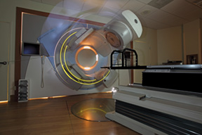
Synergy S.
Similarly, for cancers of the spine, neck, chest, lung, prostate, pancreas and liver – and for tumors in areas of the brain that are inaccessible to the Gamma Knife – the Synergy S is a highly accurate non-invasive delivery system for stereotactic radiation that combines a linear accelerator with the ability to visualize internal structure, including soft tissues, in three dimensions at the time of treatment. In this procedure, the beam of radiation moves around the patient, who is lying comfortably on a gurney on a specially fitted mesh ‘mattress’, which holds the body still during treatment. Because the radiation dose delivered by Synergy S is precisely targeted at the tumor or lesion, there’s less damage to surrounding healthy tissue. As with the Gamma Knife, the benefits of non-invasive radiosurgery and radiosurgery include no risk of blood loss, fewer complications, faster recovery and the ability to effectively treat patients who are no longer able to be treated by other methods of care. Since 2012, more than 3,000 patients have been treated, and a tremendous body of clinical research has been shared via more than 40 publications and presentations throughout the world.
Safer and more effective modalities to treat an aching back.
Today, in the hands of the highly skilled neurosurgeons at Riverside Comprehensive Neurosciences, like Dr. Dean Kostov, who have mastered its state-of-the-art surgical guidance and imaging systems, patients have a far greater likelihood of relief from debilitating back pain than ever before.
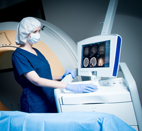
As a matter of their standard training, neurosurgeons spend a minimum of seven years learning and performing spine surgeries – including surgeries of the lumbar, cervical and thoracic spine. As a result, neurosurgeons routinely handle the depth and breadth of conditions that involve the spinal cord, the nerves and the bony structures of the spine.
The etiology of back pain can be mechanical, as in degenerative conditions like spinal stenosis, arthritis or herniated discs. It can be acquired, as in the case of scoliosis; or it can be from injuries: sprains, fractures or even osteoporosis. It can also be as a result of a primary or secondary cancer. In any case, the first and foremost goal of any spine surgery is to preserve neurological function – motor and sensory capability.
No matter how complicated the procedure, precision is absolutely critical to a safe and effective surgical result. Riverside Comprehensive Neurosciences is the only facility on the Peninsula with the capability to use both fluoroscopic and CT-based images intraoperatively. This capability, known as StealthStation, is a computer program that allows surgeons to build and visualize a 3-D model based on images obtained either from intraoperative x-rays obtained from a C-arm, or when indicated by the complexity of the pathology being treated, by intraoperative CT scans obtained from the O-arm. Because these 3-D images are more accurate than the traditional two-dimensional x-ray, the result is a quicker and more accurate operation.
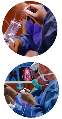 Minimally invasive surgery.
Among the reasons back surgery has such a negative reputation are the traditional methods of performing it. As open procedures, such operations required a long incision in the back that would allow the surgeon to cut down to the fascia, the fibrous tissue overlying the muscles of the spine. Once the fascia was open, the surgeon would then peel away the muscles of the spine on both sides to expose the area needing surgical intervention – be it freeing up nerves, shaving down herniated discs, or even removing entire discs to fuse the spine. The unfortunate sequelae of open surgery were muscular damage and reduced circulation. Patients faced post-operative pain and possible significant damage to the muscles surrounding the spine. Recovery was lengthy and exhausting.
In the early 1990s, researchers began exploring the surgical procedures being done successfully by general, obstetric and orthopaedic surgeons utilizing scopes, in an effort to determine whether those procedures could be adapted to spine surgery. They found that working with a scope from images on a screen resulted in a loss of three-dimensionality. They also found that a tube could work more efficiently in the narrow and crowded area of the spine. Neurosurgeons, accustomed to working with microscopes and magnifying loops, began using a tubular retractor – always with a view to maintaining spinal stability by minimizing injury to the surrounding muscles.
Today’s procedures – wider angle of attack, smaller opening.
In today’s minimally invasive spine surgery, performed by neurosurgeons at Riverside Comprehensive Neurosciences like Dr. Javier Amadeo and his colleagues, surgeons localize the target area with intraoperative x-rays, allowing them to insert a dilator, a small tube that gently nudges the muscle fibers out of their way. The ultimate result is a surprisingly wide angle of attack through a very small opening. To compensate for the inability to see spinal landmarks directly, surgeons use x-ray guidance from the C-arm imaging system, or when indicated, CT scans produced by the O-arm multidimensional platform.
Because of these exceptional visualization capabilities, even difficult and complex procedures like fusions and decompressions can be performed with these tubular retractors, using small instruments that can fit through the center of the tube.
Such procedures bring quality of life to patients who thought they had lost any opportunity to regain it – especially cancer patients whose disease has metastasized to their spine, literally crippling them. These surgeries can decompress the spinal cord, allowing patients to walk out of the hospital and into a future free of back pain.
Smaller incisions, with less bleeding and requiring shorter hospital stays, result in significantly reduced pain for the patient. But as Riverside surgeons assure their patients, it’s not the size of the incision, but what the surgeon does once inside. Their expertise at this especially complex surgery, and their mastery of the surgical techniques and tools with which they perform it, result in patients who can once again stand, sit and walk in comfort.
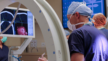
Deep brain stimulation and movement disorders.
Many of the conditions that affect the nervous system are relatively free of observable symptoms, while others produce unmistakable signs of the disease within. Two such conditions are essential tremor and Parkinson’s disease. And because the symptoms are so similar and so overtly recognizable, many people believe they’re the same condition.
But despite their seemingly similar manifestations, they are in fact very different, notes Dr. Jackson B. Salvant, a neurosurgeon who works with these patients. Essential tremor is a disorder of the nervous system that causes shaking on either side of the body. It’s most commonly a genetic disorder caused by a dominant inherited gene; that is, it tends to pass from parent to offspring. Essential tremor can affect almost any part of the body, but the shaking or trembling occurs most often in the hands — especially when attempting simple tasks like drinking from a glass, tying shoelaces, writing or shaving. Essential tremor also may affect the head, voice, arms or legs.
Parkinson’s disease is a progressive disorder of the nervous system that affects movement. It develops gradually, often starting with a barely noticeable resting tremor in just one hand. But while tremor may be the most well-known sign of Parkinson’s disease, the disorder also commonly causes a slowing or freezing of movement. Patients experience slowness of their movements, a feeling of stiffness in arms and legs, and difficulty with balance and walking. Their speech often becomes soft and mumbling. As Parkinson’s symptoms tend to worsen as the disease progresses, what began as a slight resting tremor of the hand can become very pronounced.
Significant results in controlling tremor.
No matter the etiology of the condition, patients with these conditions tend to suffer social anxiety, which especially in the case of essential tremor can exacerbate symptoms. While there is no cure for either condition, there are treatment options and techniques that can greatly improve the quality of life of these patients. But understanding the distinction between the two conditions is important, because each responds differently to different treatments.
Riverside Comprehensive Neurosciences has the only facility on the Peninsula that offers a unique treatment option to patients with either condition: deep brain stimulation, a modality that has shown significant results in controlling tremors. Prior to administering the stimulation, the patient receives mild sedation, and then under local anesthesia, is fitted with a frame similar to the Leksell frame used in Gamma Knife procedures. Once the frame is secure, the patient is awake, alert and responding to commands – enabling the surgeon to test and measure the effects of the stimulation.
Using MRI imaging and stereotactic techniques, the surgeon guides an electrical stimulation lead to a target deep within the brain in the area of the thalamus. The target areas for Essential Tremor and Parkinson’s are in relatively close proximity within the brain, but they are decidedly different in their functions and in the effects of treatment. Thus precision in reaching the appropriate target is critical.
Once a stimulating lead is precisely placed to obtain the ideal results, it is later connected to an implanted pulse generator similar to a pacemaker. The device then transmits painless electrical pulses to interrupt signals from the thalamus that may cause the tremors.
Patient selection.
Less frequently considered, but just as critical to outcomes, is the art of patient selection. Not every neurosurgical procedure is indicated for every patient with symptoms. In the age of Internet medicine, patients are more informed than ever about treatment options that are available. They are not, however, medically skilled enough to understand what will work and what will not. Additionally, some patients will bypass their family physicians to seek specialized treatment they’ve learned about when it may not, in fact, be indicated.
Patients and their referring physicians should always first seek the advice of a skilled neurologist when symptoms suggest any defect or disorder of the nervous system. Patients should be given every opportunity to try conservative forms of treatment, such as medications that may well be effective.
The medical team at Riverside Comprehensive Neurosciences insists on reserving the specialized treatment options considered herein for only those patients who will benefit the most. Each case is thoughtfully reviewed, and each procedure performed under the strictest criteria, done in coordination with other specialists.
A combination of instinct, training and skill.
It is an exquisite combination of instinct, training and skill that enables Riverside Comprehensive Neurosciences to choose the right procedure for the right patient – to perform it at the right time and with the highest standards of practice – that continue to advance this complex area of medical care to the world-class standard that the people of Greater Hampton Roads – and beyond – deserve.

